Deputy Manager — Technical; Botanic Healthcare Pvt Ltd, India
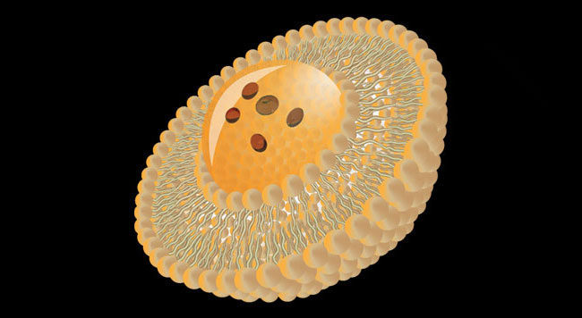
Liposomes are a form of nanocarriers that have been widely investigated for drug-delivery purposes. Liposomes have become widely used as pharmaceutical delivery carriers in numerous clinical applications. The increase in their applications has facilitated the development of analytical and technological approaches to determine their characteristics. They are composed of phospholipid bilayers which enclose a distinct aqueous space, thereby allowing encapsulation of both hydrophilic and hydrophobic compounds. The most routinely investigated parameters in liposome characterization include vesicle size and size distribution (or polydispersity), surface charge (or Zeta potential), shape and morphology, lamellarity, encapsulation efficiency, phase behaviour (or polymorphism) and in vitro release profile. A single analytical technique cannot be used to investigate the above parameters and the choice always relies on what exactly one is aiming at. Visualization analysis is an essential and powerful way for morphology studies, which allows intuitively imaging biological complex system at the molecular level. Through visualization analysis, a variety of useful data will be collected, including size distribution, bilayer organization, vesicles shape and number of lamellae. Liposome visualization data provided by microscopic techniques becomes prerequisite parameters guide the utilization of liposome for drug encapsulation.
Transmission Electron Microscopy (TEM) is an important method for the characterization of size and shape of nanoparticles as it can directly visualize single particles and even their inner architecture. The best method to visualize liposomes close to their native structure is cryo-electron microscopy, where thin films of suspensions are plunge frozen to create vitrified ice films that can be imaged directly in the electron microscope under liquid nitrogen temperature. Although subject to artifacts, negative staining TEM can also be a useful method to image liposomes, as it is faster and simpler than cryo-EM, and requires less advanced equipment. When it comes to TEM, we can understand the particle size and shape, where with cryo-TEM along with the above lamellarity can also be evaluated.
The most frequently used TEM sample preparation techniques for liposomes were drying and staining.Drying: Drying including freeze drying is the most readily accessible and widely applied method to vesicles. Typically, a drop of 2–5 μl sample is applied onto a carbon or polymer coated grid and dried for a few minutes upto several hours before imaging.
Although the most widely applied, drying is also the riskiest technique for soft materials as sometimes it aggregates upon drying and cause drying patterns, crystals and other high contrast structures to be visible. After drying, it is no longer possible to conclude whether the aggregates in the image are the result of aggregation behaviour of the sample in solution or of the sample upon drying. Even though samples exist that look the same in solution as after drying, the method of drying does not allow this distinction between what structure resulted from the sample and what structure is the result of drying.
Negative staining: Staining is better suitable for soft matter than drying. In order to preserve a sample and enhance contrast, one can use heavy metals to stain the sample. A positive stain, like iodine, ruthenium and osmium tetra oxide is a strong scattering agent that adheres to particular areas of the sample. A positive stain can change dimensions such as membrane thickness. A negative stain does not penetrate the object, but coats the surface and surroundings, obscuring the object itself and all internal structural details, and giving a foot-print like appearance. The most popular stains in soft matter are uranyl acetate (UAc) and phosphotungstic acid (PTA). Negative stain is applied to the grid straight after the sample was blotted off to prevent dehydration and interactions between the sample and the support grid. Even though the structure is much better preserved upon staining than with drying, upon staining, the sample is also dehydrated, which can cause deformation of solvent dependent structures such as lipid vesicles.
Read More...